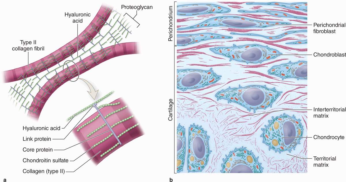
Pictures Of Cartilage Biology Diagrams Key Terms. chondroitin sulfate: An important structural component of cartilage that provides much of its resistance to compression.. connective tissue: A type of tissue found in animals whose main function is to bind other tissue systems (such as muscle to skin) or organs.It consists of the following three elements: cells, fibers, and a ground substance (or extracellular matrix).
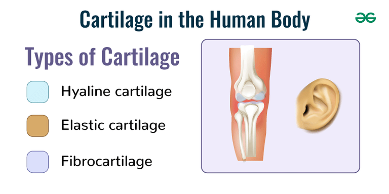
Types of cartilage. Image Source: Physiopedia. Elastic cartilage is the most flexible type of cartilage that provides support to body parts requiring bending and movement for proper function. Elastic cartilage can return to its previous shape even after enduring high pressure. Elastic cartilage is found in structures like the larynx, eustachian tubes, and external ear that require both support
Description, Anatomy, & Function Biology Diagrams
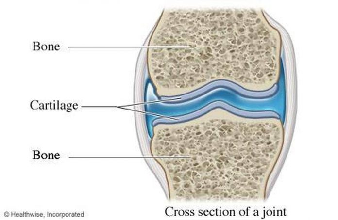
Cartilage is a connective tissue that provides support and protection for the body's joints. Cartilage located in the human body is found in joints between bones, ends of the ribs, and between the vertebrae in the spine. Cartilage is made from specialized cells called chondrocytes that produce a combination of collagen, proteoglycans, and other non-collagenous proteins.

Cartilage is a resilient and smooth type of connective tissue.Semi-transparent and non-porous, it is usually covered by a tough and fibrous membrane called perichondrium.In tetrapods, it covers and protects the ends of long bones at the joints as articular cartilage, [1] and is a structural component of many body parts including the rib cage, the neck and the bronchial tubes, and the
Cartilage: Anatomy, histology, types and functions Biology Diagrams
Cartilage, connective tissue forming the mammalian embryonic skeleton prior to bone formation and persisting in parts of the human skeleton into adulthood. It is composed of a dense network of collagen fibers embedded in a gelatinous ground substance. Learn more about the structure and function of cartilage.