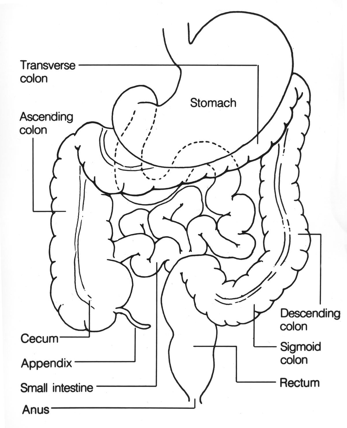
Some facts about colonoscopy Biology Diagrams The colon (large intestine) is the distal part of the gastrointestinal tract, extending from the cecum to the anal canal. It receives digested food from the small intestine, from which it absorbs water and electrolytes to form faeces. Anatomically, the colon can be divided into four parts - ascending, transverse, descending and sigmoid. These sections form an arch, which encircles the small
Your large intestine has three parts: the colon, rectum and anus. Each part does specific things to keep food waste moving through your large intestine. The colon. Your colon has five parts that work to process food waste and move waste to your rectum. Those parts are: Cecum: This is the first part of your colon. It's about 3 inches (8 cm) long.
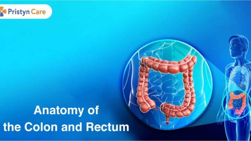
The Rectum: Anatomy and 3D Illustrations Biology Diagrams
Anatomy of Colon and Rectum. The entire colon is about 5 feet (150 cm) long, and is divided into five major segments. The rectum is the last anatomic segment before the anus.. The ascending and descending colon are supported by peritoneal folds called mesentery.. The right colon consists of the cecum, ascending colon, hepatic flexure and the right half of the transverse colon. The rectum is the final segment of the large intestine that connects the colon to the anus. It stores fecal matter produced in the colon until the body is ready to eliminate the waste through the process of defecation. Anatomy. The rectum is a hollow muscular tube about 8 inches (20 cm) in length and 2.5 inches in diameter at its widest point.
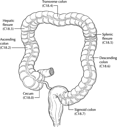
The job of the sigmoid colon is to solidify stool before it enters the rectum and anal canal for excretion. It does this by contracting, and the increased pressure moves the stool. Your rectum is at the end of your large intestine, a long, continuous tube that includes your colon, rectum and anus. Your rectum makes up the last 6 inches or so, just before it turns into the anal canal. This is the last stop on your food's journey through your gastrointestinal (GI) tract before it exits. As food waste passes from your

Colon anatomy: Pictures, features, and function Biology Diagrams
10.1055/b-0038-166135 1 Surgical Anatomy of the Colon, Rectum, and AnusSanthat Nivatvongs, Philip H. Gordon, and David E. Beck Abstract This chapter will review the surgical anatomy of the colon and rectum, including general configuration, course relations, peritoneal coverings, and ileocecal valve in the colon, and peritoneal relations, fascial attachments, fascia propria, Waldeyer's fascia Anatomy of the Colon and Rectum Richard L. Drake Jennifer M. McBride Introduction Fig. 1. The components of the large intestine, which consists of the cecum, the appendix, and the ascending, transverse, descending, and sigmoid colons, the rectum and the anal canal. (From Drake RL, Vogl W, Mitchell AWM, et al. Gray's Atlas of Anatomy.…